OPTG (2D) and CBCT (3D) provide complementary ways to examine the mouth and jaws.
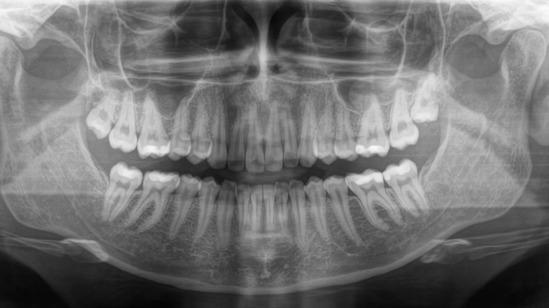
OPTG, also known as panoramic dental imaging or an orthopantomogram, is a Finnish invention. It is an important diagnostic tool and is used, for example, when checking for dental infection before joint replacement surgery. It provides a two-dimensional image of the jaws, jaw joints, maxillary sinuses, nasal cavity and teeth. Infections around tooth roots can often be identified, and digital images can be adjusted for examination. The image also shows the anatomy around wisdom teeth and nerves, additional teeth, milk teeth and areas of infection. A radiology report is prepared and entered into Kanta. According to the latest information provided to the clinic, dental X-ray images will become available in Kanta in 2029.
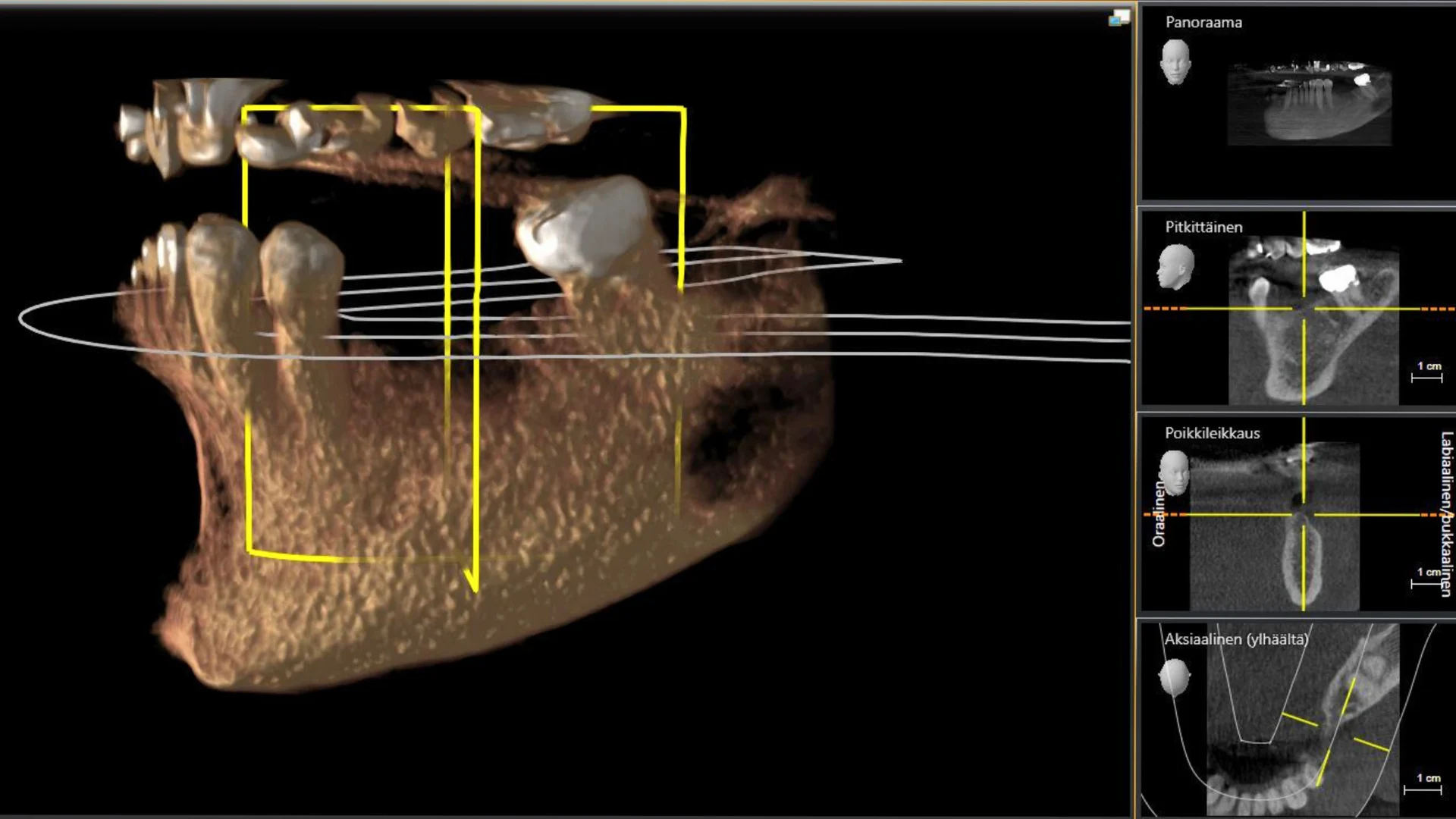
CBCT, or cone beam computed tomography, uses a computer to reconstruct a three-dimensional X-ray image of the area examined. It is used to investigate root fractures, additional root canals, nerve positions and bone anatomy. Low-dose imaging with a small field of view is also used for implant planning. Before surgery, a virtual implant-supported crown is positioned in relation to the bone and bite. A surgical guide is then made to transfer the plan to the procedure and guide implant placement. Planning in advance helps avoid pitfalls and surprises.
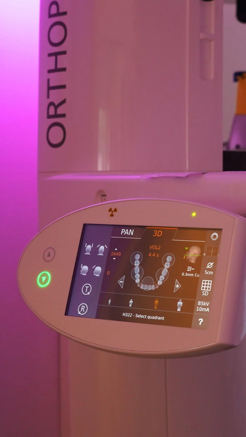
MedNiemi also accepts imaging referrals from other dentists. Kela reimbursement for CBCT imaging requires a referral from a specialist dentist. Our dental X-rays have always been digital. Our panoramic and CBCT unit is the Dentsply Sirona Orthophos SL.